As Brain Activity Increases, What Happens To Heart Rate?
Abstract
Many people experience balmy stress in modern society which raises the need for an improved understanding of psychophysiological responses to stressors. Middle rate variability (HRV) may be associated with a flexible network of intricate neural structures which are dynamically organized to cope with diverse challenges. HRV was obtained in thirty-three healthy participants performing a cerebral task both with and without added stressors. Markers of neural autonomic command and neurovisceral complexity (entropy) were computed from HRV time serial. Based on private anxiety responses to the experimental stressors, two subgroups were identified: anxiety responders and non-responders. While both vagal and entropy markers rose during the cerebral job alone in both subgroups, simply entropy decreased when stressors were added and exclusively in anxiety responders. We conclude that entropy may be a promising marking of cognitive tasks and acute mild stress. It brings out a new fundamental question: why is entropy the only marker affected by mild stress? Based on the neurovisceral integration model, we hypothesized that neurophysiological complexity may be contradistinct by mild stress, which is reflected in entropy of the cardiac output betoken. The putative role of the amygdala during mild stress, in modulating the complication of a coordinated neural network linking brain to center, is discussed.
Introduction
Many people feel stress in mod social club, which can strongly influence their mental and physical well-being. Curt-term physiological and psychological responses to stress are healthy regulations; yet, more prolonged exposure or inadequate responses to stressors can lead to depletion of resources and can be a source of chronic diseases1,ii.
Nosotros can undoubtedly chronicle the concept of stress to the works of Walter Cannon3, who first described short-term physiological changes giving ascension to the fight-or-flying behavioral responses. After on, Hans Selye4, past studying various physiological stressors, observed consistent effects on the organisms leading him to define stress as "the non-specific response of the body to whatsoever demand upon it". From the works of Masonfive, most researchers have reconsidered the model of Selye when dealing with psychological stressors, since they are tied to the way nosotros translate situations or events6,vii,viii. This points out that stress is rather a highly individual feel based on the transaction betwixt an individual and its environment5,6. Every bit a affair of fact, the stress reactivity of the organism relies on the personal perception, interpretation and appraisal made nigh the stressors7,8, which are themselves driven by diverse individual determinants (e.thou. personality, socio-economic status…)9,ten,11,12. The estimation of unpredictability, novelty and uncontrollability of a given situation, only also its character of social evaluative threat, are the iv factors highlighted to imply a stress response from the trunkfive,xiii.
When 1 individual interprets a situation as stressful, there is a need for the body to trigger acute changes that serve the mobilization of energy and resources to coordinate an acceptable response3,4,5,14,15,16,17,xviii. This response requires the activation of the sympathetic-adrenal-medullary and the hypothalamic-pituitary adrenal axesxvi,17,18. The associated secretion of catecholamines and glucocorticoids allows for brusk-term changes in blood pressure and centre rate, making the cardiovascular system peculiarly sensitive to the stress furnishings14.
The analysis of heart rate variability (HRV, or RR interval time serial) provides information on changes in the tone of vagal (parasympathetic) and sympathetic control of the autonomic nervous organisation that triggers most of short-term cardiovascular modulations19,20. HRV analyses are believed to provide valuable and relevant markers of acute stress21. Connections between the autonomic regulations and the primal nervous system have been best-selling since the 90's22, and farther evidenced by neuroimaging studies23,24,25,26,27. Through the cardinal autonomic network (CAN), the encephalon exerts a tight control on a wide diverseness of the body's regulations (e.g. viscera, hormonal secretion) that are disquisitional for flexibility and goal-directed behavior22. The CAN components are distributed over a number of interconnected neural structures and parallel pathways managing multiple inputs and outputs22,28. This intricate network is at the origin of the circuitous control of heart rate dynamics driven past vagal and sympathetic modulationsxx. From this view point, HRV reveals more only a healthy cardiac part, only also how the encephalon achieves flexible control and adaptive regulationsxix, allowing us to explore coordinated heart-brain interactions through the concept of neurovisceral integration22,29.
HRV measures have allowed to appraise fundamental nervous processes, such every bit attentional control, emotional regulation and knowledge. These assessments mainly focused on time- and frequency-domain analyses of HRV time series21,24,25,28. A dandy deal of attention has been paid to the tonic vagal control28,thirty,31,32, defined as the vagal control under resting-land weather condition, where a authorisation of vagal ability has been linked to an effective prefrontal tonic activity25,28,29,33. Under normal conditions, the prefrontal activeness consists of the constant inhibition of subcortical structures such as amygdala25,34. Much less is known on the acute (phasic31) response to cognitive challenges. Confusing results take been reported, evidencing either a blunted23,35,36,37 or an enhanced31,38 vagal response. Hence, while the relationship between vagal function and cerebral processes seems obvious25,28, it appears to considerably depend on the context31. Park et al.31 suggested an expected ascent of vagal response due to one's cocky-regulatory effort. All the same, this vagal response could exist blunted by an unfavorable context.
When the context is perceived as stressful, information technology is widely acknowledged that the circuitry linking the prefrontal cortex and the amygdala plays a key function in the stress-related changes in behavior and peripheral physiological reactivity through the autonomic control39,40. Feet is known to impair the recruitment of prefrontal control mechanisms and to disrupt this circuitry, which can lead to an amygdaloid hyper-responsivity41.
It follows from the above that physiological stress furnishings are initiated in the brain, involve a complex neural organization spanning multiple cerebral structures and intricate autonomic integration, that ultimately impacts cardiac regulations. The literature increasingly finds an interesting correspondence between the circuitous organization of such neurophysiological systems and the complexity of the time series obtained from their indicate outputs42,43. Recently, a link has been shown between complexity markers extracted from HRV fourth dimension serial and cognition, mood and state anxiety44,45. Mainly, entropy (a computation of signal irregularity) has the capacity to reflect the complexity of control systems, like the 1 decision-making heart rate42. In Information theory, entropy describes the rate of information that is created past a stochastic source of data. This is also seen in physiological complexity, where the functioning of healthy systems is complex by nature, and a shift toward simplicity is associated with a loss of flexibility and adaptability. As an illustration, cardiac entropy has been shown to vanish with aging and pathology42,46,47. This suggests that an entropy-based approach may add together significant value to the understanding of circuitous neural organization and heart-brain interactions challenged past stressful and/or cognitive conditions44,45.
In the present study, 30-three healthy participants were asked to perform a cognitive job in unlike contexts: with and without added stressors, while heart rate dynamics were recorded. While vagal and sympathetic markers were assessed past classic time- and frequency-domain analyses of HRV, the novelty lies in the utilize of entropy to detect complexity in coordinated heart-brain interactions, in clan with stress and cerebral task.
Methods
Population
The study group consisted of 33 good for you volunteers (age: 35.6 ± 13.9 years, 19 women). All the participants gave their written informed consent to participate in the present study in accordance with the principles of the Declaration of Helsinki. The study was approved past the institutional review board of the kinesthesia of sport sciences, Academy of Bordeaux, France. All the experiments respected the principles set up past the CNIL (Commission Nationale de 50'Informatique et des Libertés), the CPP (Comité de Protection des Personnes), and the ARS (Agence Régionale de Santé). Participants were recruited amongst university students and employees: all of whom have had a academy pedagogy or were undertaking one. Participants were eligible to participate if they did not present prior cardiovascular illnesses (e.yard. arrhythmia, center failure), severe inflammation (e.g. arthritis) or psychological disorders (e.g. burn-out syndrome). Furthermore, they did not accept any medication influencing the cardiovascular arrangement (e.g. antidepressant, antipsychotic, antihypertensive, psychotropic). Participants were asked to avert ingestion of booze and caffeinated beverages for the 12 hours preceding each period and to abstain from heavy physical activeness the 24-hour interval earlier the series of experiments.
Protocol
The experimental protocol was designed as three successive situations of a duration of 8 min (as determined by pre-testing), separated past most 10 min during which participants filled out psychological questionnaires. During each situation, participants were seated in front of a computer, breathing at spontaneous rate, while RR interval time series were recorded as described below. The first situation was systematically the reference state of affairs (Ref.), where participants were asked to relax, watching an emotionally neutral documentary picture show about whales (L'odyssée des baleines à bosses, Ross Isaacs, Stan Esecson, 2022). During the following two situations, occurring in a randomized order, they performed a cognitive task without stressors (CT) or with additional stressors (CT + S). Randomization was performed using the formula =alea() in a spreadsheet, where participants were attributed a randomized number that adamant the order of the situations.
Heart charge per unit recordings and analyses
RR interval time series were directly obtained with ±1 ms accurateness from a bipolar electrode transmitter belt Polar H7 (Polar, Finland) fitted to the breast of the participant and continued to an iPod (Apple, Cupertino CA, USA) via Bluetooth. A smartphone awarding (HRV Logger®) was used to continuously store RR intervals transmitted by the Polar device. The reliability and accuracy of this Polar device take been previously demonstrated, through comparisons with electrocardiography (ECG) measurements48,49,50. Over eight min periods, nearly 400–500 successive RR intervals were obtained, the verbal length of the RR interval time series depending on the average individual middle charge per unit.
The nerveless RR interval fourth dimension serial were exported to Matlab (Matworks, Natick MA, Us) for farther analysis, using available functions and custom-designed routines. Initially, the raw data were inspected for artifacts20. Occasional ectopic beats (irregularity of the center rhythm involving extra or skipped heartbeats, e.g. extrasystole and consecutive compensatory break), were visually identified and manually replaced with interpolated next RR interval values.
RR interval fourth dimension series were analyzed in time-domain and in frequency-domain. The average RR duration, RMSSD (square root of the mean of the sum of the squared differences between adjacent normal RR) too as power vs. frequency relation were computed, afterward 4 Hz (regular) resampling using cubic spline interpolation. Power was calculated in fixed bands between 0.04 Hz to 0.15 Hz for the low frequencies (LF, associated with sympathetic activity) and between 0.15 Hz and 0.four Hz for the high frequencies (HF, associated with vagal activeness) after Fourier transform of the resampled RR interval time seriestwenty. The ratio LF/HF was calculated, as an index of the sympathovagal resttwenty.
For the purpose of the present study, complexity in the neurovisceral control of the center was assessed by computing entropy in RR interval fourth dimension series, using a recently improved routine: refined composite multiscale entropy (RCMSE)51. This method is based on multiscale entropy (MSE) and its variant, composite multiscale entropy (CMSE), which were adult to assess complexity in physiological output signals, based on sample entropy over several scales42. At each level of resolution (scale), MSE yields a value that reflects the mean rate of creation of data. The overall degree of complexity of a signal is then calculated past integrating the values obtained for a pre-defined range of scales. As discussed by Wu et al.51, RCMSE improved the accuracy of MSE (and CMSE) by reducing the probability of inducing undefined entropy. For the analysis of short time series (equally in the present study), RCMSE is strongly recommended51.
Briefly, the RCMSE algorithm consists of the following procedures (see detailed method in51):
- (i)
The RR interval time series is coarse grained using overlapping windows to obtain the representation of the original time series on unlike time scales τ. Overlapping windows allow for grand coarse-grained series at each calibration factor of τ.
- (2)
At each scale gene of τ, the number of matched vector pairs \({n}_{k,\tau }^{m+one}\) and \({n}_{k,\tau }^{grand},\) is calculated for all \((chiliad)\,\tau \) coarse-grained series, with \(m\) (here, \(m=two\)) corresponding to the sequence length considered. This calculation refers to the probability that segments (vectors) of \(grand\) samples that are similar, remain like when the segment length increases to \(m+1\).
- (3)
The RCMSE at a scale factor of τ is provided as follows, with r corresponding to the tolerance for matches. In the nowadays study, \(r=0.15\) of the standard deviation of the initial time series:
$$RCMSE(x,\,\tau ,\,m,\,r)=-\,\mathrm{ln}(\frac{{\sum }_{yard=ane}^{\tau }{northward}_{g,\tau }^{m+i}}{{\sum }_{k=1}^{\tau }{n}_{k,\tau }^{m}})$$
(1)
In our conditions, RCMSE was assessed over the range of scales one to 3, larger scales existence overlooked due to the risk of unreliable results51,52. The cardiac entropy index was calculated from the area under the curve of entropy vs. scale (using the trapezoidal rule) for scales 1, 2 and three as illustrated in Fig. 1.
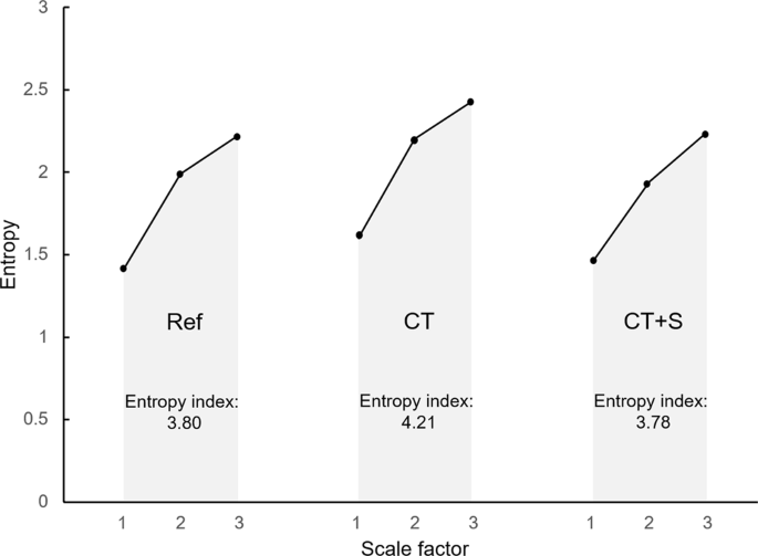
Typical curves of entropy vs. scales in the 3 experimental situations for one participant. Following a fibroid-graining procedure on the RR interval time serial, refined composite multiscale entropy (RCMSE) was assessed over the scale 1 to 3. The entropy index was computed for each experimental situation, Ref., CT and CT + S, by calculating the expanse under the curve. Ref., reference state of affairs; CT, cognitive task situation; CT + S, cerebral task situation under stress.
Cognitive task
During both the CT and CT + S situations, participants had to perform a nonverbal cerebral task comprising memorization, mental calculation and logic questions. This task was created using the East-Prime software (Psychology Software Tools Inc., Pittsburgh, PA). The participants answered by typing on the computer's keyboard. A full of 23 items were presented in CT and 31 items in CT + South (more items in the same 8 min total duration because express time to answer an item was part of the stress induction). Despite 23 vs. 31 items, the content of the items remained the aforementioned (memorization, mental calculation and logic questions) in each situation CT and CT + Due south.
Stress induction
To induce stress during CT + S, stressors were chosen based on a meta-analysis of 208 laboratory stress studiesxiii. It was shown that physiological responses to stressors are exacerbated when a participant is exposed to a combination of an uncontrollable environs and a social-evaluative threat. Therefore, each question in CT + S was displayed for a predefined time not controllable past the participant. In add-on, a visual feedback was displayed when the participant gave a incorrect answer. Tertiary, two other persons were present in the room with the participant and acted as an circumspect and evaluative audition. Finally, a variety of sound disturbances (e.yard. crowd noise) were played continuously during the 8 min situation.
Psychological questionnaires
Participants filled out two questionnaires after each state of affairs in order to appraise their state of feet and cognitive workload levels. The Spielberger'south Country-trait feet inventory (STAI) was administered to the participants53. The STAI state anxiety scale consists of 20 questions that evaluate the current state of anxiety past using items that measure subjective feelings of apprehension, tension, nervousness and worry. The use of NASA chore load index (NASA-TLX) immune a cocky-assessed mensurate of workload based on half-dozen components: mental need, physical demand, temporal need, performance, effort, and frustration level54. Participants were asked to evaluate each component on a calibration, and the weight of each component was so assessed before computing a global index, which is the weighted boilerplate of said components.
Statistical assay
All statistical procedures were conducted past use of XLSTAT (Addinsoft, 2022, XLSTAT statistical and information analysis solution, Long Island, NY, USA). Quantitative measurements are expressed every bit mean ± standard deviation.
The group size was determined by power calculation (GPower 3.1.nine.2) based on our preliminary data obtained during pre-testing (α mistake probability: 0.05, power 0.eight) hateful entropy value, iii.61; standard deviation, 0.47; mean difference 8% [0.289]. This resulted in northward = 34 (actually 33 participants were ultimately recruited) for one experimental group (Cohen's d event size: 0.35). No formal power calculation was performed for time- and frequency-domain markers.
Repeated measures analysis of variance (ANOVA) testing with the mail service hoc Tukey correction was used to appraise the effects of experimental situations on cardiac markers, state anxiety and cognitive workload scores. The variables satisfied the conditions of normality, tested with Shapiro-Wilk examination. Unpaired t-test was used when state anxiety was compared between subgroups. Pearson correlation calculations were used to examine the relationship between changes in country anxiety score and cardiac entropy index. A value of p < 0.05 was considered to signal statistical significance.
Results
Land anxiety scores
While the country feet scores exhibited a modest range beyond participants both in Ref. and in CT (respectively: twenty–44 and twenty–48), in contrast, the range was greater when stressors were added in the CT + S situation (22–76). When analyzed in more detail, this greater range in CT + S immune u.s.a. to identify participants with markedly dissimilar behaviors and led u.s.a. to divide the whole population into two subgroups: anxiety responders and anxiety not-responders. The quantitative criterion for subgroup constitution was the stressors-induced changes in individual country anxiety scores, observed when comparing CT and CT + S - as illustrated in Fig. 2. Those individuals exhibiting more a twenty% increase in their anxiety score due to stressors were included in the subgroup of anxiety responders (north = 20). The remaining people were included in the subgroup of feet not-responders (northward = xiii).
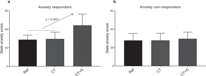
State anxiety score for each experimental situation. Feet responders are represented in (a) and anxiety not-responders in (b). Ref., reference situation; CT, cognitive task situation; CT + S, cognitive task state of affairs nether stress. Error confined represent the standard difference.
This subdivision did not generate any divergence in averaged anxiety score in Ref. state of affairs (29 ± 5 in responders and 28 ± 8 in not-responders). The execution of a cognitive task (CT) did not result in a change in state anxiety in any subgroup. As expected, considering information technology was the benchmark for subgroup subdivision, feet score in response to stressors (Fig. 2) increased in anxiety responders (+32 ± 8%, p < 0.0001) just not in anxiety non-responders (+6 ± 10%, ns).
Cognitive workload scores
When analyzing changes in cognitive workload score in response to CT and CT + Southward, we observed consistent typical beliefs in anxiety responders and feet not-responders (Fig. 3). The score increased in response to a cognitive task (+119 ± 76%, p < 0.0001, in responders and +118 ± 151%, p = 0.0002, in non-responders) and increased over again when stressors were added (+48 ± 46%, p < 0.0001, in responders and +33 ± 22%, p = 0.0002, in non-responders).
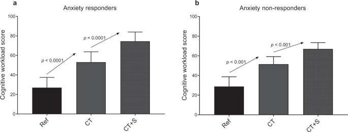
Cerebral workload score for each experimental situation. Anxiety responders are represented in (a) and anxiety non-responders in (b). Ref., reference state of affairs; CT, cognitive chore situation; CT + Due south, cerebral task situation under stress. Error bars represent the standard difference.
Cardiac autonomic markers
In both subgroups, the hateful RR did not change in whatsoever situation (Table 1). RMSSD increased between Ref. and CT in each subgroup (p = 0.026 in feet responders and p = 0.025 in anxiety non-responders). Among frequency-domain markers of HRV, only HF power increased between Ref. and CT in non-responders (p = 0.005). Both results are associated with vagal enhancement in CT. No change was observed in other autonomic markers (LF power, LF/HF, Table ane). Information technology is worth noting that none of these autonomic markers changed in response to stressors (CT + Southward, Tabular array ane).
In contrast with the to a higher place archetype cardiac markers in time-domain and frequency-domain, the entropy marker revealed the physiological impact of stressors during the cognitive task (Tabular array 1, Fig. 4). Farther, changes in cardiac entropy brought valuable information about stress when analyzed in relation to the higher up-mentioned subjective ratings of state feet and cognitive workload. While the entropy index increased in CT, both in anxiety responders and non-responders (+11 ± 19%, p = 0.026, in responders and +8 ± 10%, p = 0.028, in non-responders), only anxiety non-responders were able to maintain a loftier level of entropy in presence of stressors (CT + S). In contrast, entropy dropped in anxiety responders (−8 ± 10%, p = 0.005, Fig. iv).
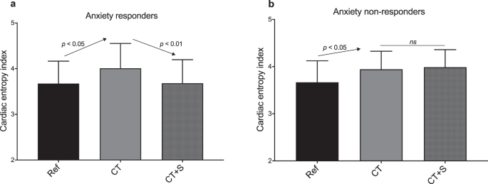
Cardiac entropy alphabetize for each experimental state of affairs. Anxiety responders are represented in (a) and anxiety non-responders in (b). Ref., reference situation; CT, cognitive task situation; CT + S, cognitive task situation under stress. Mistake bars represent the standard departure.
A deeper analysis of individual responses strengthened the link between entropy and feet. As shown in Fig. five, when stressors were added to CT, individual changes in land anxiety level were correlated with private changes in cardiac entropy: the greater anxiety, the greater drop in entropy.
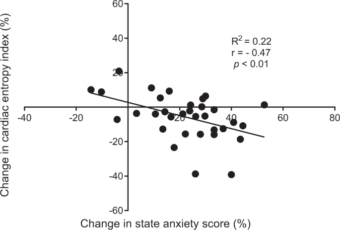
Correlation analysis betwixt change in land feet score and change in cardiac entropy index, from the cognitive task (CT) to the cognitive task under stress (CT + S). Changes are expressed in percentage difference.
Discussion
Previous studies accept described top-down middle-brain interactions through a complex network involving the autonomic nervous control of the centre rate28. Here, we have hypothesized that cognitive-induced neural modulations would reflect in signal complication of RR interval time series, which could be degraded past stress42,55. A ready of vagal, sympathetic and entropy markers was obtained during cognitive job and balmy stress situations. The main finding was that both vagal tone and entropy increased during the cognitive task, but entropy alone reflected psychophysiological responses to balmy stress. In addition, the correlation between entropy changes and anxiety responsiveness shed new light on the intricate neurophysiological functions and how they are impacted by stress.
Because that the cardiac response induced by a cerebral task should differ from that induced past stressors25,31,32,56, a challenge in the present study was to distinguish psychological stress from cognitive workload57,58. The cognitive workload is described as the mental toll to operate a task54 and has been shown to increase with working memory tasks and during problem solving59. In our conditions, this was reflected in the ascension of cognitive workload estimated by the participants in CT (Fig. 3). All participants, irrespective of their subgroup (anxiety responders or anxiety not-responders), perceived the cognitive workload without experiencing an increment in feet (Fig. two).
The rise in cognitive workload was concomitant to a heightened vagal influence on cardiac control (Table one). Several studies take linked the parasympathetic modulations of the cardiac rhythm by the vagus nerve to cognitive processes, pointing out an alphabetize of how strongly top-down appraisals, mediated by cortical-subcortical pathways, shape brainstem activity and autonomic responses24,25,31. Within the framework of the neurovisceral integration model, most studies have focused on the vagal function at remainder, associated with the concept of tonic vagal control25,56,threescore,61. Hither, we instead focused on the acute (phasic) vagal response, related to baseline-to-task changes28,31, which has been shown to depend on the context31. In our conditions, the observed heightened vagal control in every participant is in understanding with a better attentional control31.
In the nowadays study, two subgroups were described amongst participants, anxiety responders and anxiety non-responders (Fig. ii). This distinction was motivated by the observation that state anxiety response to stressors (CT + S vs. CT) was highly heterogeneous, and the volition to understand the psychophysiological significant of the differences. Anxiety is defined as a negative emotional response to threatening circumstances which is associated with physiological stress arousal62,63. It is common to find different anxiety patterns when people are facing stressful situations13,45, considering there is no mutual way to react to stressful and challenging environmental exposure64. By definition, a stressor is a stimulus that triggers a physiological response when a potential threat is perceived by the brain. This perception depends on intrinsic individual factors65, such as personalitynine, socioeconomic status10, personal history or stored memory66. As illustrated, a same stressful state of affairs (CT + South) was interpreted either as innocuous or as a potential threat from one participant to another, equally shown by various individual state feet. This different perception of stressors, influenced sensory inputs and their respective processing, which led to the well-described variability in physiological responses13,67,68,69.
As a primary finding here, when stressors were added to the cognitive task (CT + Southward), no additional effect was reflected in vagal markers, whatever the subgroup (Table one). Information technology has been recently claimed that not only vagal merely as well sympathetic control could interfere in the relationship between cerebral processes and autonomic cardiac regulations30. This is particularly relevant when studying non-resting weather condition, specially those consisting of stress induction. In our weather, the cardiac sympathetic mark (LF ability) did non rise in CT or in CT + South (Table i). In sum, the analyses of cardiac markers in time-domain (RMSSD) and frequency-domain (HF, LF, LF/HF) were unable to detect stress responses whatsoever the participant contour, be it an anxiety responder or non-responder. It is worth noting that a balmy stress was nether investigation hither, which could explain the absence of change in center rate (run across mean of RR intervals in Tabular array one), and vagal or sympathetic tones, even in anxiety responders.
As a fundamental point in the present work, the benefit of a complexity marker to explore neurovisceral integration during balmy stress was shown. There is recent evidence that complication emerges as a promising framework to analyse cardiac-ending signals as a reliable picture of intricate cortical, subcortical and peripheral interactions within the Can. This prove is reinforced when studying noesis and stress that claiming a flexible organization coordination44,45. Although it is acknowledged that the very mechanisms at the origin of complexity in fourth dimension series are scarcely identified, physiological complexity has been associated with healthseventy and an elevated capacity to adapt to an e'er-changing environment. Among complication markers, entropy is defined as the main alphabetize able to inform about the charge per unit of information product71, a rate that is heightened in a coordinated system. Notably, in our weather, cardiac entropy increased during the cognitive task (CT) in each subgroup (Fig. four), concomitantly with the ascension in cognitive workload. This likely reflects a coordinated, flexible and robust neural network taking place for optimal perceptual and cognitive functioning. Recent neuroimaging studies evidenced that the degree of complexity in cerebral BOLD (Blood Oxygenation Level-Dependent) signals is positively correlated with cognitive processes such equally attention, memory and verbal fluency72. Therefore, the rise in cardiac entropy observed in our participants might be an indicator of enhanced heart-brain interactions.
Past using a multiscale entropy approach, we showed a decrease in entropy in CT + S state of affairs in anxiety responders, whereas feet non-responders maintained the heightened entropy gained during CT (Fig. 4). Nosotros draw a parallel with the decrease in cardiac entropy recently showed during university examinations45. This leads us to conclude that using a robust entropy-based method of HRV, entropy is a relevant marking of stress-induced changes in eye-encephalon interactions, even in mild stress conditions. The link observed here, between stress-related anxiety and breakdown in cardiac complexity during a cerebral job, finds support in contempo functional imaging of the brain. Neuroimaging has evidenced a reduction in both prefrontal cortex and anterior cingulate cortex activities together with an increase in amygdala activity, associated with anxiety and stress41,73,74,75. In improver, anxiety reduced top-down control and connectivity between these structures, thus creating a bias towards related responses41,74. Such impairments might exist at the origin of the loss of cardiac entropy observed in the present written report in anxiety responders. Cardiac entropy could reflect central and autonomic regulations and consequently could reveal the alteration of heart-brain connectivity wherein the amygdala activity is involved. The amygdala-driven disruption in cortical-subcortical interactions may provide a kind of information overflow that impairs coordination between multiple interacting components. Typically, a degraded coordination in the neurophysiological arrangement has been shown to reverberate in the complication of cardiac-catastrophe point outputs42. In this scenario, a less adaptive and flexible cardiac autonomic control results from the issue of anxiety on the amygdala, which is reflected in the cardiac signal entropy nether mild stress. A potentially of import consequence of this could exist that exploring cardiac complexity is critical for the exploration of psychophysiological manifestations of stress.
Despite appealing outcomes, the electric current written report is not without limitations. While an adequate total number of participants was achieved, as assessed past pre-hoc ability-calculation, the results obtained in our male and female person participants were analyzed birthday. However, a sexual dimorphism has been evidenced in psychophysiological responses to stress and anxiety44, so that a greater number of female participants should make it possible to explore the sex-related nature of complex heart-brain interactions operating during cerebral tasks with stressors. Additionally, part of our assay led us to distinguish two subgroups among our participants: anxiety responders and anxiety non-responders, with respective sample size: northward = xx and n = 13. Given the pre-hoc power-calculation, at that place is a risk of fake negative, especially in non-responders (n = 13) when the hypothesis is rejected that entropy does not drop with stressors. Yet, it is worth noting that at an private level (due north = 33), we also observe a significant correlation (p < 0.01, Fig. 5) between change in entropy and anxiety responsiveness. Thus, the main conclusion, that a degraded cardiac entropy reflects the neurovisceral integrated response to anxiety during a cognitive task receives strong support. Finally, all the participants had university level of education and were accepted to performing challenging cognitive tasks, which requires farther investigation involving people without academy teaching before our results could be generalized.
Determination
Nosotros found show that cardiac entropy changes concomitantly with acute responses to cognitive load and stress. While cardiac entropy could be a marking of enhanced complexity and acceptable cocky-regulation during a cognitive job, a degraded entropy in cardiac betoken outputs might reverberate an overflow of neural information. This overflow might be due to an amygdala-induced disruption in the cortical-subcortical processing in broken-hearted people. While information technology is obvious that entropy-based approaches should not replace spectral analysis of HRV – and their chapters to brand a distinction betwixt vagal and sympathetic responses –, exploring complication in the neurophysiological control of eye charge per unit likely adds significant value to our understanding of neurophysiological operation in association with the feet-targeted function of the amygdala.
References
-
Lupien, S. Well Stressed: Manage Stress Before It Turns Toxic. (John Wiley & Sons, 2022).
-
Yaribeygi, H., Panahi, Y., Sahraei, H., Johnston, T. P. & Sahebkar, A. The impact of stress on body function: A review. EXCLI J 16, 1057–1072 (2017).
-
Cannon, W. B. The wisdom of the body. (W W Norton & Co, 1932).
-
Selye, H. Stress without Distress. in Psychopathology of Human Accommodation (ed. Serban, One thousand.) 137–146, https://doi.org/10.1007/978-1-4684-2238-2_9 (Springer The states, 1976).
-
Mason, J. W. A review of psychoendocrine research on the sympathetic-adrenal medullary organization. Psychosom Med 30(Suppl), 631–653 (1968).
-
Lazarus, R. Southward. & Folkman, S. Stress, apraisal, and coping (1984).
-
Liu, J. J. W., Ein, Northward., Gervasio, J. & Vickers, K. The efficacy of stress reappraisal interventions on stress responsivity: A meta-analysis and systematic review of existing show. PLOS Ane 14, e0212854 (2019).
-
Raymond, C., Marin, M.-F., Juster, R.-P. & Lupien, S. J. Should we suppress or reappraise our stress?: the moderating role of reappraisal on cortisol reactivity and recovery in salubrious adults. Feet Stress Coping 32, 286–297 (2019).
-
Soliemanifar, O., Soleymanifar, A. & Afrisham, R. Human relationship betwixt Personality and Biological Reactivity to Stress: A Review. Psychiatry Investig 15, 1100–1114 (2018).
-
Boylan, J. M., Cundiff, J. M. & Matthews, K. A. Socioeconomic Status and Cardiovascular Responses to Standardized Stressors: A Systematic Review and Meta-Assay. Psychosom Med 80, 278–293 (2018).
-
Bibbey, A., Carroll, D., Roseboom, T. J., Phillips, A. C. & de Rooij, S. R. Personality and physiological reactions to acute psychological stress. Int J Psychophysiol 90, 28–36 (2013).
-
Liu, J. J. Reframing the Private Stress Response: Balancing our Knowledge of Stress to Improve Responsivity to Stressors., https://doi.org/10.17605/OSF.IO/CTD4V (2019).
-
Dickerson, South. S. & Kemeny, K. Due east. Astute stressors and cortisol responses: a theoretical integration and synthesis of laboratory research. Psychol Balderdash 130, 355–391 (2004).
-
McEwen, B. Due south. Stress, accommodation, and disease. Allostasis and allostatic load. Ann. Due north. Y. Acad. Sci. 840, 33–44 (1998).
-
Juster, R.-P., Perna, A., Marin, K.-F., Sindi, S. & Lupien, Southward. J. Timing is everything: Anticipatory stress dynamics amid cortisol and claret force per unit area reactivity and recovery in healthy adults. Stress 15, 569–577 (2012).
-
Lupien, Due south. J. et al. Across the stress concept: Allostatic load–a developmental biological and cognitive perspective. in Developmental psychopathology: Developmental neuroscience, Vol. ii, 2nd ed 578–628 (John Wiley & Sons Inc, 2006).
-
Murison, R. Chapter 2 - The Neurobiology of Stress. in Neuroscience of Pain, Stress, and Emotion (eds. al'Absi, M. & Flaten, M. A.) 29–49, https://doi.org/ten.1016/B978-0-12-800538-5.00002-9 (Academic Printing, 2022).
-
Dhabhar, F. S. The Short-Term Stress Response – Mother Nature's Mechanism for Enhancing Protection and Operation Under Atmospheric condition of Threat, Challenge, and Opportunity. Front Neuroendocrinol 49, 175–192 (2018).
-
Ernst, K. Center-Charge per unit Variability—More than than Heart Beats? Front Public Health v (2017).
-
Heart rate variability. Standards of measurement, physiological estimation, and clinical use. Task Force of the European Society of Cardiology and the Northward American Guild of Pacing and Electrophysiology. Eur. Centre J. 17, 354–381 (1996).
-
Kim, H.-G., Cheon, E.-J., Bai, D.-S., Lee, Y. H. & Koo, B.-H. Stress and Heart Charge per unit Variability: A Meta-Assay and Review of the Literature. Psychiatry Investig fifteen, 235–245 (2018).
-
Benarroch, E. E. The central autonomic network: functional system, dysfunction, and perspective. Mayo Clin. Proc. 68, 988–1001 (1993).
-
Gianaros, P. J., Van Der Veen, F. K. & Jennings, J. R. Regional cerebral blood period correlates with centre catamenia and loftier-frequency heart menstruum variability during working-memory tasks: Implications for the cortical and subcortical regulation of cardiac autonomic action. Psychophysiology 41, 521–530 (2004).
-
Lane, R. D. et al. Neural correlates of heart rate variability during emotion. Neuroimage 44, 213–222 (2009).
-
Thayer, J. F., Ahs, F., Fredrikson, 1000., Sollers, J. J. & Wager, T. D. A meta-analysis of heart rate variability and neuroimaging studies: implications for heart rate variability as a marker of stress and health. Neurosci Biobehav Rev 36, 747–756 (2012).
-
Yoo, H. J. et al. Brain structural concomitants of resting land middle rate variability in the young and old: evidence from two independent samples. Encephalon Struct Funct 223, 727–737 (2018).
-
de la Cruz, F. et al. The human relationship between heart rate and functional connectivity of brain regions involved in autonomic control. Neuroimage 196, 318–328 (2019).
-
Thayer, J. F. & Lane, R. D. Claude Bernard and the heart-encephalon connectedness: further elaboration of a model of neurovisceral integration. Neurosci Biobehav Rev 33, 81–88 (2009).
-
Thayer, J. F. & Lane, R. D. A model of neurovisceral integration in emotion regulation and dysregulation. J Affect Disord 61, 201–216 (2000).
-
Giuliano, R. J., Gatzke-Kopp, L. M., Roos, L. East. & Skowron, Eastward. A. Resting sympathetic arousal moderates the clan between parasympathetic reactivity and working memory performance in adults reporting loftier levels of life stress. Psychophysiology 54, 1195–1208 (2017).
-
Park, G., Vasey, Grand. W., Van Bavel, J. J. & Thayer, J. F. When tonic cardiac vagal tone predicts changes in phasic vagal tone: the part of fear and perceptual load. Psychophysiology 51, 419–426 (2014).
-
Thayer, J. F. & Sternberg, E. Beyond heart rate variability: vagal regulation of allostatic systems. Ann. N. Y. Acad. Sci. 1088, 361–372 (2006).
-
Beauchaine, T. P. & Thayer, J. F. Eye rate variability as a transdiagnostic biomarker of psychopathology. Int J Psychophysiol 98, 338–350 (2015).
-
Barbas, H. & García-Cabezas, M. Á. How the prefrontal executive got its stripes. Curr. Opin. Neurobiol. twoscore, 125–134 (2016).
-
Bernardi, L. et al. Effects of controlled breathing, mental activity and mental stress with or without verbalization on heart rate variability. J. Am. Coll. Cardiol. 35, 1462–1469 (2000).
-
Mathewson, Thousand. J. et al. Autonomic predictors of Stroop performance in young and middle-aged adults. Int J Psychophysiol 76, 123–129 (2010).
-
Overbeek, T. J. 1000., van Boxtel, A. & Westerink, J. H. D. M. Respiratory sinus arrhythmia responses to cerebral tasks: effects of chore factors and RSA indices. Biol Psychol 99, i–14 (2014).
-
Segerstrom, S. C. & Nes, L. S. Heart rate variability reflects self-regulatory strength, effort, and fatigue. Psychol Sci xviii, 275–281 (2007).
-
LeDoux, J. The emotional brain, fear, and the amygdala. Prison cell. Mol. Neurobiol. 23, 727–738 (2003).
-
Arnsten, A. F. T. Stress weakens prefrontal networks: molecular insults to higher cognition. Nat. Neurosci. 18, 1376–1385 (2015).
-
Bishop, South. J. Neurocognitive mechanisms of anxiety: an integrative account. Trends Cogn. Sci. (Regul. Ed.) 11, 307–316 (2007).
-
Costa, G., Goldberger, A. Fifty. & Peng, C.-K. Multiscale entropy assay of biological signals. Phys Rev E Stat Nonlin Soft Matter Phys 71, 021906 (2005).
-
Lipsitz, L. A. Dynamics of stability: the physiologic basis of functional health and frailty. J. Gerontol. A Biol. Sci. Med. Sci. 57, B115–125 (2002).
-
Young, H. & Benton, D. We should be using nonlinear indices when relating middle-charge per unit dynamics to cognition and mood. Scientific Reports 5, 16619 (2015).
-
Dimitriev, D. A., Saperova, E. V. & Dimitriev, A. D. Country Feet and Nonlinear Dynamics of Middle Rate Variability in Students. PLOS Ane 11, e0146131 (2016).
-
Chen, C.-H. et al. Complication of Center Rate Variability Can Predict Stroke-In-Evolution in Acute Ischemic Stroke Patients. Scientific Reports v, 17552 (2015).
-
Lin, Y.-H. et al. Reversible center rhythm complication impairment in patients with main aldosteronism. Scientific Reports 5, 11249 (2015).
-
Weippert, Yard. et al. Comparison of 3 mobile devices for measuring R-R intervals and centre rate variability: Polar S810i, Suunto t6 and an convalescent ECG organisation. Eur. J. Appl. Physiol. 109, 779–786 (2010).
-
Vasconcellos, F. V. A. et al. Middle charge per unit variability assessment with fingertip photoplethysmography and polar RS800cx as compared with electrocardiography in obese adolescents. Blood Printing Monit 20, 351–360 (2015).
-
Giles, D., Draper, N. & Neil, W. Validity of the Polar V800 heart rate monitor to measure RR intervals at rest. Eur J Appl Physiol 116, 563–571 (2016).
-
Wu, S.-D., Wu, C.-W., Lin, Southward.-G., Lee, Thousand.-Y. & Peng, C.-K. Assay of complex time series using refined composite multiscale entropy. Physics Letters A 378, 1369–1374 (2014).
-
Gow, B. J., Peng, C.-M., Wayne, P. Yard. & Ahn, A. C. Multiscale Entropy Assay of Center-of-Pressure Dynamics in Man Postural Control: Methodological Considerations. Entropy 17, 7926–7947 (2015).
-
Spielberger, C. D., Gorsuch, R., Lushene, E. R., Vagg, P. & Jacobs, A. G. Manual for the State-Trait Feet Inventory (Class Y1 – Y2). IV (1983).
-
Hart, S. Yard. & Staveland, 50. E. Development of NASA-TLX (Task Load Alphabetize): Results of Empirical and Theoretical Inquiry. in Advances In Psychology (eds. Hancock, P. A. & Meshkati, North.) 52, 139–183 (North-Holland, 1988).
-
Delignieres, D. & Marmelat, V. Fractal fluctuations and complication: current debates and future challenges. Crit Rev Biomed Eng 40, 485–500 (2012).
-
Park, G., Vasey, G. W., Van Bavel, J. J. & Thayer, J. F. Cardiac vagal tone is correlated with selective attention to neutral distractors under load. Psychophysiology fifty, 398–406 (2013).
-
Cinaz, B., Arnrich, B., Marca, R. & Tröster, G. Monitoring of Mental Workload Levels During an Everyday Life Role-piece of work Scenario. Personal Ubiquitous Comput. 17, 229–239 (2013).
-
Setz, C. et al. Discriminating Stress From Cerebral Load Using a Wearable EDA Device. IEEE Transactions on Information technology in Biomedicine 14, 410–417 (2010).
-
Berka, C. et al. EEG correlates of job engagement and mental workload in vigilance, learning, and retention tasks. Aviat Space Environ Med 78, B231–244 (2007).
-
Jennings, J. R., Allen, B., Gianaros, P. J., Thayer, J. F. & Manuck, Due south. B. Focusing neurovisceral integration: cognition, centre charge per unit variability, and cognitive blood flow. Psychophysiology 52, 214–224 (2015).
-
Hansen, A. L., Johnsen, B. H. & Thayer, J. F. Vagal influence on working retentiveness and attending. Int J Psychophysiol 48, 263–274 (2003).
-
Meijer, J. Stress in the relation between trait and state anxiety. Psychol Rep 88, 947–964 (2001).
-
Eysenck, One thousand. West., Derakshan, N., Santos, R. & Calvo, 1000. Chiliad. Anxiety and cerebral functioning: attentional control theory. Emotion seven, 336–353 (2007).
-
Carroll, D. Wellness Psychology: Stress, Behaviour and Affliction. 1st ed., (1992).
-
Novais, A., Monteiro, S., Roque, S., Correia-Neves, M. & Sousa, Due north. How age, sexual activity and genotype shape the stress response. Neurobiol Stress half-dozen, 44–56 (2016).
-
Cicchetti, D. Resilience under weather of extreme stress: a multilevel perspective. World Psychiatry nine, 145–154 (2010).
-
Carnevali, L., Koenig, J., Sgoifo, A. & Ottaviani, C. Autonomic and Encephalon Morphological Predictors of Stress Resilience. Front Neurosci 12 (2018).
-
Lupien, S. J., McEwen, B. S., Gunnar, M. R. & Heim, C. Effects of stress throughout the lifespan on the brain, behaviour and cognition. Nat. Rev. Neurosci. 10, 434–445 (2009).
-
Ouellet-Morin, I. et al. Enduring effect of babyhood maltreatment on cortisol and eye rate responses to stress: The moderating role of severity of experiences. Dev. Psychopathol. 1–12, https://doi.org/ten.1017/S0954579418000123 (2018).
-
Goldberger, A. L. et al. Fractal dynamics in physiology: alterations with affliction and aging. Proc. Natl. Acad. Sci. Usa 99(Suppl i), 2466–2472 (2002).
-
Pincus, S. Yard. & Goldberger, A. L. Physiological time-serial analysis: what does regularity quantify? Am. J. Physiol. 266, H1643–1656 (1994).
-
Yang, A. C. et al. Complexity of spontaneous BOLD action in default mode network is correlated with cognitive office in normal male elderly: a multiscale entropy assay. Neurobiol. Aging 34, 428–438 (2013).
-
Gianaros, P. J. et al. Individual differences in stressor-evoked blood pressure reactivity vary with activation, book, and functional connectivity of the amygdala. J. Neurosci. 28, 990–999 (2008).
-
Bishop, S., Duncan, J., Brett, K. & Lawrence, A. D. Prefrontal cortical function and feet: controlling attention to threat-related stimuli. Nat. Neurosci. 7, 184–188 (2004).
-
Shin, Fifty. One thousand. et al. An fMRI written report of anterior cingulate function in posttraumatic stress disorder. Biol. Psychiatry 50, 932–942 (2001).
Acknowledgements
The authors give thanks Lauren Smith for reviewing the manuscript.
Author information
Affiliations
Contributions
E.B., L.Yard.A. and V.D.-A. wrote the main manuscript text. E.B., H.One thousand., 5.50.-N., E.Yard. and Five.D.-A. designed the experiment. E.B. executed the experiment. E.B., 50.1000.A., P.G., 5.L.-North., Eastward.G. and V.D.-A. analyzed the data. All authors reviewed the manuscript.
Corresponding writer
Ethics declarations
Competing interests
The authors declare no competing interests.
Additional information
Publisher'due south note Springer Nature remains neutral with regard to jurisdictional claims in published maps and institutional affiliations.
Rights and permissions
Open up Access This article is licensed under a Creative Commons Attribution iv.0 International License, which permits use, sharing, adaptation, distribution and reproduction in whatsoever medium or format, as long as y'all requite appropriate credit to the original author(s) and the source, provide a link to the Creative Commons license, and indicate if changes were made. The images or other tertiary party textile in this article are included in the article'due south Artistic Commons license, unless indicated otherwise in a credit line to the material. If material is non included in the commodity'due south Creative Commons license and your intended utilise is not permitted by statutory regulation or exceeds the permitted employ, y'all will need to obtain permission directly from the copyright holder. To view a copy of this license, visit http://creativecommons.org/licenses/by/iv.0/.
Reprints and Permissions
Nigh this article
Cite this commodity
Blons, E., Arsac, 50.One thousand., Gilfriche, P. et al. Alterations in eye-encephalon interactions under mild stress during a cognitive task are reflected in entropy of heart charge per unit dynamics. Sci Rep 9, 18190 (2019). https://doi.org/10.1038/s41598-019-54547-7
-
Received:
-
Accepted:
-
Published:
-
DOI : https://doi.org/x.1038/s41598-019-54547-7
Further reading
Comments
By submitting a comment you concord to bide by our Terms and Customs Guidelines. If you discover something abusive or that does not comply with our terms or guidelines please flag it as inappropriate.
As Brain Activity Increases, What Happens To Heart Rate?,
Source: https://www.nature.com/articles/s41598-019-54547-7
Posted by: schaferevess1985.blogspot.com


0 Response to "As Brain Activity Increases, What Happens To Heart Rate?"
Post a Comment